Nucleic Acids
DNA Structure
The structure of DNA was determined in 1953, by Watson and Crick. It was confirmed by x-ray crystallography, which is a method that rely on x-rays being scattered by a crystalline piece of material. The pattern of scattering gives clues about the structure.
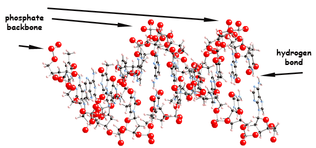
 Figure 1 - Structure of DNA (it is shown in the movie below in different positions)
 Figure 2 - Close up view of figure 1, showing details of the hydrogen bonds, on the G-C base pair.
The chemical composition was already known. Biochemical studies had shown that DNA is composed of phosphate, sugar and nitrogenated bases. An important clues to the structure was provided by the discovery that equal numbers of the bases A and T were present, and the same for the bases C and G. That suggested that these bases are paired, what was later confirmed. The importance of the phosphate backbone and the hydrogen bonds was known, but there were doubts about the position of the phosphate backbone, which was thought to be at the center of the molecule, and later it was confirmed to be on the outside.
The helical structure was a good guess, considering the fact that Linus Pauling had already discovered that the secondary structure of certain proteins was a helix. The hydrogen bonds are essential for the structure.
Figure 2 (above) shows the hydrogen bonding between G, on the left, and C, on the right. Notice that this is a triple hydrogen bonding. The other base pair, A and T, only have a double hydrogen bonding. It also shows the phosphate on the far right hand side (check colour coding of atoms).

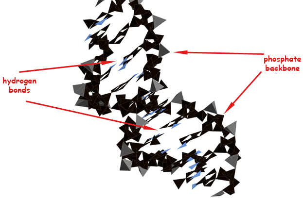
Figure 3 - Polyhedral representation
The polyhedral representation of the DNA molecule, above, makes clear the position of structural details like the phosphate backbone and the hydrogen bonds. The terahedra on the backbone represent phosphate groups, which are tetrahedral.
Movie - The structure of DNA from different point of views (colour coding above).
© Ricardo Esplugas de Oliveira -2020

